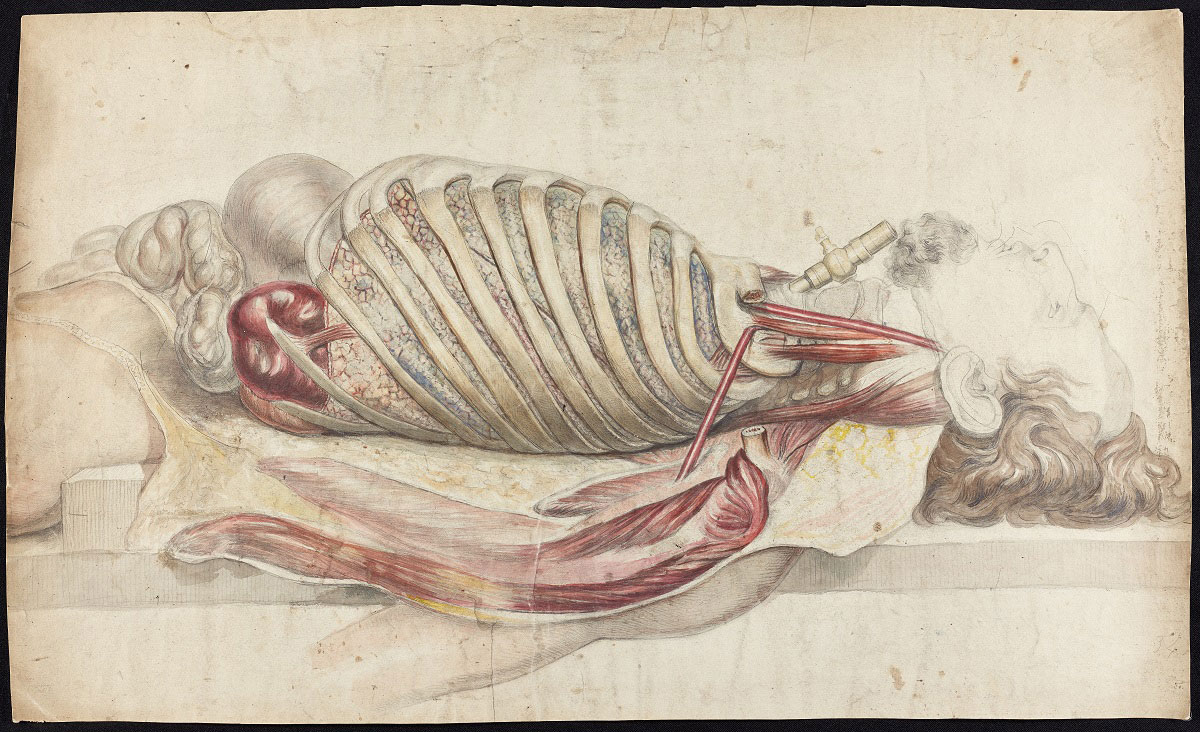
For a few weeks, there’s an exhibition about what lies beneath the skin of the average human being.
As a species we are fascinated by the contents of our complex and fragile bodies, and physicians, scientists, surgeons, artists and printers have developed tools and techniques, from ancient woodcuts to contemporary three-dimensional imaging, in order to identify and understand what is hidden inside the human form.

The results, while aimed at improving heathcare, are also often quite beautiful and at times utterly gross, yet still presented beautifully.
A short-lived exhibition has just opened at the Royal College of Physicians, next to Regent’s Park, and although it’s only open Mon-Fri, they’re having two late night openings for people stuck at work during normal hours.
The exhibition, ‘Under the skin: illustrating the human body’ explores the art and science of anatomical illustration from the medieval world to the present day. Using rarely seen books, artworks and objects from the Royal College of Physicians’ collections, the displays examine the themes of illustrating, opening, mapping, knowing and treating the human body.
Amongst the many highlights of the exhibition is a complete edition of Andreas Vesalius’ De humani corporis fabrica libri septem, published in Basel in 1543. One of the most famous books in the history of medicine and of art, it depicts the human body with a level of detail, accuracy and creative flair completely unknown before.
Nearby, a 17th-century work produced in London shows a flayed man standing as if still alive, holding up his own skin, the features of his face still clearly visible on the ghost-like surface.
Equally as shocking is a brightly coloured engraving by Jacques Gautier d’Agoty from 18th century Paris. In it two heads are shown, once more as if still living but this time lying closely together as though in a bed. At first sight they appear to be drawn with the accuracy and sympathy of a portrait. On second glimpse it’s clear that their skulls and facial features have been dissected.

A remarkable image created in Persia (present day Iran) in 1656, uses feathered lines to indicate the widespread of the nerves throughout the body, various colours tracing the different branches and routes of the fibres.
From Victorian Scotland comes a startling photographic image of 1893 showing a horizontal cross-section of the human brain, seeming to visually presage the scans of the modern age.
The exhibition at the Royal College of Physicians is open until 15th March, and is open Mon-Fri 9am to 5pm.
There are two late nights though, on Thur 7th Feb and 7th March until 8pm — with curator tours at 6pm.
Entry is free.
The exhibition will return in an extended run from early October 2019.